Your journey to optimal
wellness begins here.
At Altus Medical Imaging, we understand that accurate and precise
diagnostics are the cornerstone of effective healthcare.
With a team of dedicated professionals and the latest imaging equipment,
we provide you with unparalleled insight into your health.
Medical Imaging Services
Our dedicated team of experts is committed to visualising your health with precision and care, ensuring you receive the best possible medical attention.
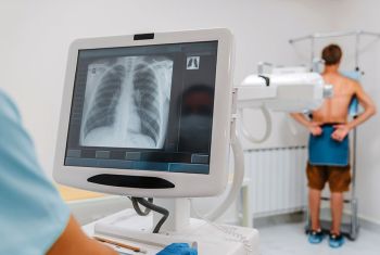
X-Ray
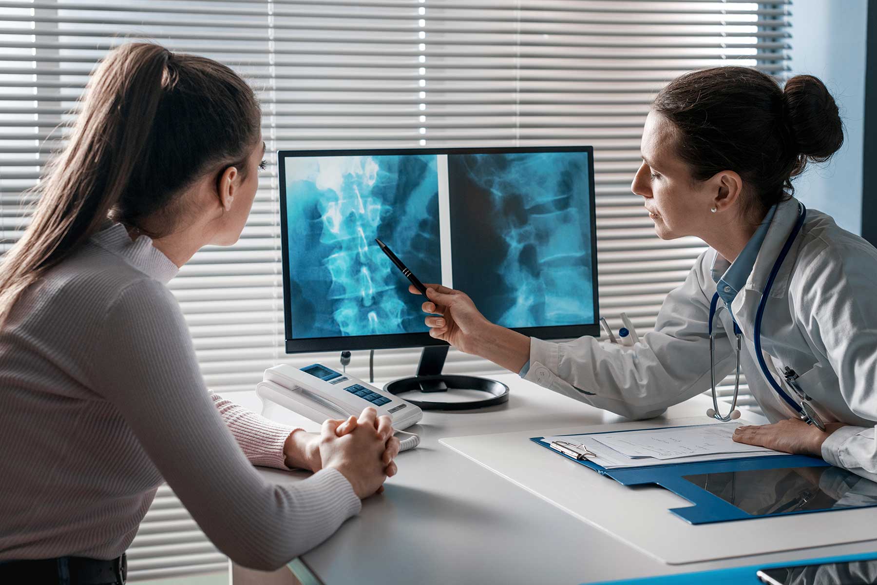
An x-ray image, or radiograph, is produced when a very small amount of ionising radiation passes through the body and strikes a sensitive detector placed on the other side of the body. X-ray imaging can be used to assess various body parts, commonly bones, joints, the lungs and the abdomen.

Ultrasound
Ultrasound is a safe and widely used imaging technique producing detailed pictures of the body in real time, using high frequency sound waves which are produced by a special ultrasound probe, called a transducer. Ultrasound has no known harmful effects and can be used to image a variety of conditions including pregnancy, gallstones and varicose veins. Ultrasound can also be used to measure blood flow through vessels, and this is called a doppler or duplex scan.
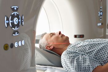
CT – Low Dose
Low radiation dose computed tomography (CT) uses low levels of radiation to help diagnose and monitor a wide array of conditions. Using the CT scanner and a powerful computer, we can build 3D images showing the soft tissues, bones and blood vessels, and see parts of the body, which are difficult to view by any other method. Most CT examinations are simple, fast and pain free. The CT scanning machine combines x-rays with computer technology to produce more detailed, cross-sectional images of the body. Wherever possible, PRC performs low radiation dose computer tomography.

3D Mammography
A mammogram is a low dose x-ray of the breast. Mammograms are performed for two major reasons. Firstly, in patients with breast symptoms, to detect a possible cause for their symptoms (a diagnostic mammogram), and secondly, to detect early signs of breast cancer in patients who do not have breast symptoms (a screening mammogram).
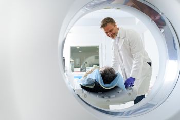
MRI
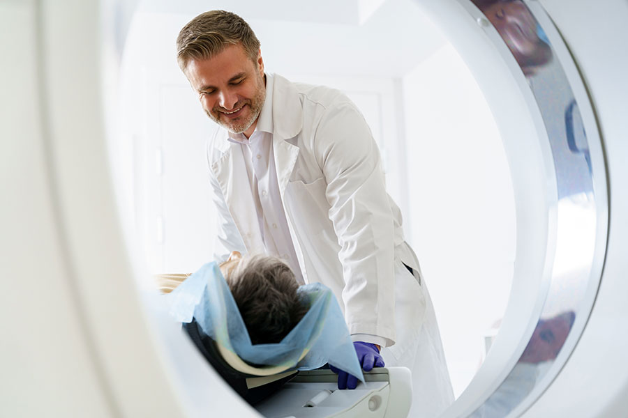
Magnetic resonance imaging (MRI) is a safe, painless and powerful diagnostic imaging test. MRI technology is very complex but essentially uses a strong magnetic field and radio waves to produce exquisite images of many of the body’s internal structures. MRI is a very safe test because it does not use radiation to collect the images.
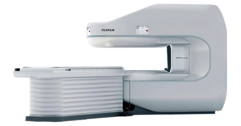
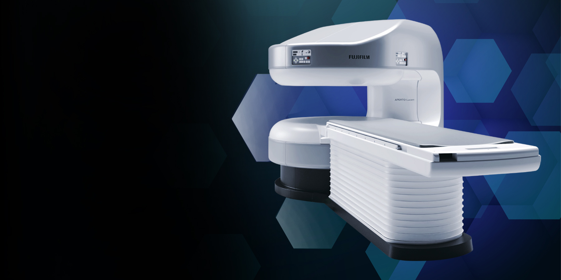
APERTO Lucent Open MRI
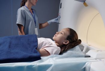
Paediatric Imaging
Our scans and radiological examinations are performed and interpreted by paediatric specialists with decades of vast clinical experience working at one of Australia’s top tertiary children’s hospitals.
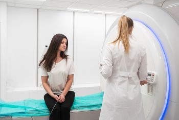
Women’s Imaging
Altus provides a range of pregnancy ultrasounds including dating scans, Nuchal Translucency, morphology and 4D scans.
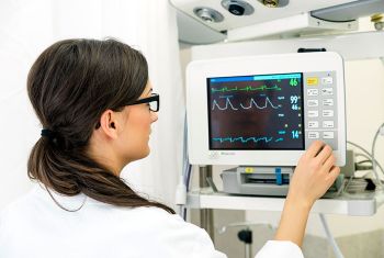
Cardiac Imaging
CT coronary angiogram uses a powerful X-ray machine to produce images of the heart and its blood vessels. The test is used to diagnose a variety of heart conditions.
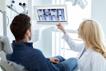
Dental Imaging
Using specialised equipment to assess the jaw and teeth, for example, impacted teeth, pre-implant assessment, orthodontic assessment, and assessment for jaw and face surgery.
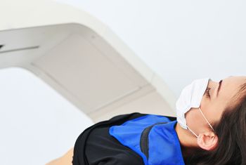
Dexa (BMD)
Dual energy x-ray absorptiometry (DEXA) is used to measure bone mineral density (BMD). Low bone density or osteoporosis is a major determinant of fracture risk and is common, particularly in women following menopause. Loss of bone density may also be accelerated in conditions affecting the liver and thyroid gland and by certain medications, in particular steroids. Measurement of the forearm may also be undertaken if required.

Injections & Biopsies
We are proud to offer a range of injections for pain relief or to assist the muscle, joint and tendon healing process.
Facet joint injection
Nerve root sleeve injection and epidural
Injections – Muscle, joint or tendon
Cortisone
Platelet Rich Plasma

Euflexxa
Euflexxa is used to relieve joint pain caused by osteoarthritis of the knee, hip, shoulder and ankle. It is an injectable synthetic version of hyaluronan, the key ingredient of synovial fluid found naturally in the joint space. With osteoarthritis, the normal synovial fluid becomes dysfunctional, leading to friction and inflammation. Euflexxa may reduce pain and reinstate joint flexibility and mobility, enhancing the overall quality of life for patients.
Our Doctors at Altus
A Wealth of Expertise
We’re proud to have a team of highly skilled and experienced doctors who are dedicated to your well-being. Our medical professionals are committed to providing the highest quality of care and accuracy in diagnostic imaging.
Discover Your Closest Clinic
to assist. Find out the nearest clinic for your convenience.
About Altus Medical Imaging
Where Clarity and Compassion Meet Innovation
Whether you require routine screenings, specialised diagnostics, or comprehensive imaging solutions, our mission is to empower you and your healthcare provider with the visual information needed to make informed decisions about your health.
